• Skip to main content
  • Welcome
    • Accreditation
    • IWAS 2019 – Video Modules
    • Recording Inaugural Online Class of March 2021
  • Registration
  • Program
  • 2025 IAS Case Competition
  • Our Supporters
  • Contact Us
  • Past Conferences
    • Photo Gallery
  • Unaccredited Sessions

IAS

International Arrhythmia School

Home » Past Conferences » 2015

2015

PosterCME AccreditationWorkshopsFacultySupporting OrganizationsLocationAbstractsPresentation DownloadsPhotos

Program

CME Accreditation

12th International Winter Arrhythmia School was approved for the following credits by Continuing Professional Development, Faculty of Medicine, University of Toronto:

Royal College of Physicians and Surgeons of Canada – 17.5 Section 1 credits
The American Medical Association – 17.5 AMA PRA Category 1 credits
The College of Family Physicians of Canada – 4.0 Mainpro-M1 credits

Through an agreement between the Royal College of Physicians and Surgeons of Canada and the American Medical Association, physicians may convert Royal College MOC credits to AMA PRA Category 1 Credits™. Information on the process to convert Royal College MOC credit to AMA credit can be found at www.ama assn.org/go/internationalcme.

12th International Winter Arrhythmia School was endorsed by:

Workshops

Building on the success of previous conferences, our workshops offered specialized instruction for nurses, EP technicians, residents, fellows, and allied professionals.

We deployed world-class faculty to engage in discussions and expect, as always, fierce educational argument.

  • Telemetry Workshop (Level 1-3)
  • ECG Workshop
  • Ultrasound Guided Vascular Hands-on Workshop
  • The Subcutaneous ICD Hands-on Workshop
  • Ontario Association of Cardiologists Workshop

International Winter Arrhythmia School Founder and Chair: Dr. Eugene Crystal

12th Annual Hosting Faculty

  • Dr. Eugene Crystal, Chair, Toronto, Ontario
  • Claudia Bucci, Toronto, Ontario
  • Dr. Ilan Lashevsky, Toronto, Ontario
  • Dr. Edgar Hockmann, Toronto, Ontario
  • Dr. Darren Kagal, Toronto, Ontario
  • Dr. Bruce lubelsky, Toronto, Ontario
  • Dr. David Newman, Toronto, Ontario
  • Dr. Mohammed Shurrab, Toronto, Ontario
  • Dr. Irving Tiong,Toronto, Ontario
  • Suzette Turner, NP, Toronto, ON

12th Annual International Faculty

  • Dr. David Burkhardt, Austin, USA
  • Dr. Martin C. Burke, Chicago, USA
  • Dr. Fermin C. Garcia, Philadelphia, USA
  • Dr. Lior Gepstein, Haifa, Israel
  • Dr. Hamid Ghanbari, Ann Arbor, USA
  • Dr. Gregory Golovchiner, Petah Tikva, Israel
  • Dr. Hiroshi Nakagawa, Oklahoma City, USA
  • Dr. William Spear, Oak Lawn, USA
  • Dr. Tamas Szili-Torok, Rotterdam, Netherlands
12th Annual National Faculty

  • Dr. Guy Amit, Hamilton, Ontario
  • Dr. Sean Balmain, Toronto, Ontario
  • Dr. Brian Baker, Toronto, Ontario
  • Dr. Adrian Baranchuk, Kingston, Ontario
  • Dr. Sacha Bhatia, Toronto, Ontario
  • Dr. David Birnie, Ottawa, Ontario
  • Debra Campbell, RN, Kingston, Ontario
  • Dr. Michael Hartleib, Peterborough, Ontario
  • Dr. Jeff Healey, Hamilton, Ontario
  • Dr. Amir Janmohamed, Toronto, Ontario
  • Dr. Peter Leong-Sit, London, Ontario
  • Dr. Bhavanesh Makanjee, Toronto, Ontario
  • Dr. Carlos Morillo, Hamilton, Ontario
  • Dr. Grish M. Nair, Ottawa, Ontario
  • Dr. Pablo Nery, Ottawa, Ontario
  • Dr. Doug Ng, Toronto, Ontario
  • Dr. Vladislav Ovchinnikov Vladislav, Toronto, Ontario
  • Dr. Arnold Pinter, Toronto, Ontario
  • Dr. Kieran Quinn, Kingston, Ontario
  • Dr. Sherryn Rambihar, Toronto, Ontario
  • Dr. James Swan, Toronto, Ontario
  • Dr. Anthony Tang, London, Ontario
  • Dr. Derek Yung, Toronto, Ontario

Organizers  Banafsheh Arouny & Seddigheh Baktash

Supporting Organizations

We are grateful to the following partners for their generous support of last year’s International Winter Arrhythmia School:

12th International Winter Arrhythmia School is endorsed by:

2015 Sponsors:


Location

The 12th International Arrhythmia School was hosted at the Westin Trillium House at Blue Mountain in Collingwood, Ontario. Blue Mountain is a ski resort located approximately 90 minutes by car outside of Toronto. 220 Gord Canning Drive (Formerly Mountain Drive), Blue Mountains Ontario, L9Y 0V9, Canada Phone: (705) 443-8080

Abstracts

Obstructive Sleep Apnea as a Predictor of Post Coronary Artery Bypass Graft Atrial Fibrillation: A Systematic Review and Meta-analysis
Authors: Amro Qaddoura, Conrad Kabali, Doran Drew, Erik M van Oosten, Kevin A Michael, Damian P Redfearn, Christopher S Simpson, Adrian Baranchuk
Queen’s University – School of Medicine

Background: Post coronary artery bypass graft atrial fibrillation (PCAF) is associated with increased morbidity, mortality, and system costs. Few studies explored obstructive sleep apnea (OSA) as a risk factor for PCAF. We aimed to systematically review and synthesize the evidence associating OSA with PCAF.

Methods: We conducted a search in MEDLINE, Embase, Google Scholar, Web of Science, abstracts, conference proceedings, and reference lists until June 2014. Eligible studies were English, conducted in humans, and assessed OSA with polysomnography or a validated questionnaire. Two reviewers independently selected studies, with disagreement resolved by consensus. Piloted forms were used to extract data and assess risk of bias.

Results: Five prospective cohort studies were included (n = 642). There was agreement in study selection (κ 0.89, 95% confidence interval [CI] 0.75 to 1.00). OSA was associated with a higher risk of PCAF (odds ratio [OR] 1.86, CI 1.24 to 2.80; p = 0.003; I2 = 35%). We conducted three subgroup analyses. The associations increased for data that used polysomnography to assess OSA (OR 2.34, CI 1.48 to 3.70), when severe OSA was included from one study (OR 2.59, CI 1.63 to 4.11), and when adjusted analyses were pooled (OR 2.38, CI 1.57 to 3.62; p

Discussion: We found OSA to increase the odds of PCAF by approximately two-fold. Despite the multifactorial risk for PCAF, we are confident in the validity of the unadjusted analyses given their similarity to the adjusted analysis. We did not identify any investigations assessing the effectiveness of OSA management strategies in reducing PCAF and other complications in this population, making it an appealing future research topic.

Conclusions: OSA was shown to be a strong predictor of PCAF. Future research efforts should aim to assess the effectiveness of OSA identification and management strategies in reducing PCAF and other complications in this population.

Feasibility and Safety of Using Mechanic and Magnetic Robotic Systems for EGM-Guided AF Ablation with Near Zero Radiation: A Pilot Study
Authors: Atsuhiko Yagishita, MD; Harish Manyam, MD; Samer De Oliveira, MD; Alvaro Manrique, MD; Michael Arredondo, MD; Harikrishnan Santhakumari, MD; Ivan Cakulev, MD; Judith Mackall, MD; Mauricio Arruda, MD
Harrington Heart and Vascular Institute, University Hospitals Case Medical Center, Case Western Reserve University, School of Medicine, Cleveland, Ohio, USA
Abstract:
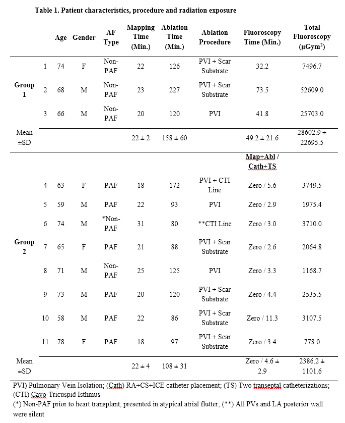
Purpose: This pilot study was designed to assess the feasibility and safety of using the Stereotaxis and the AmigoTM Remote Catheter System (RCS) as a non-fluoroscopic approach for EGM-guided atrial fibrillation (AF) ablation.
Methods: This prospective study enrolled 11 consecutive patients presenting for radiofrequency (RF) catheter ablation of AF. Group 1. The initial 3 patients underwent 3-D electroanatomic voltage mapping while creating LA and PV antra geometry. This was obtained by roving a custom, bidirectional multi-electrode circular catheter remotely via the Amigo RCS. Subsequently, PV electrical isolation (PVI) and adjunctive ablation was performed by remote RF delivery via the Stereotaxis magnetic system. This phase was designed to allow the operator to become familiar with the Amigo remote navigation and fluoroscopy was allowed as needed. Group 2. The subsequent 8 patients underwent the same procedure to test the following hypothesis: “A fully robotic EGM-guided AF ablation can be performed without or minimal radiation exposure”.
Results: All 11 patients underwent successful robotic EGM-guided PVI, scar substrate and CTI ablation, as needed using the Stereotaxis and the Amigo RCS systems. In all patients in Group 2, LA and RA mapping (22 ± 4 min.) and ablation (108 ± 31 min.) were performed without fluoroscopic. The mean fluoroscopic time and total radiation exposure (μGym2) was 49.2 ± 21.6 vs. 4.6 ± 2.9 min., and 28602.9 ± 22695.5 vs. 2386.2 ± 1101.6 μGym2 for Group 1 and Group 2, respectively (P < 0.001). There were no complications and AF recurred in 1 patient during short-term follow-up (99-126 days).
Conclusion: This pilot study suggests that a fully robotic EGM-guided AF ablation and adjunctive ablation can be performed safely and with minimal radiation exposure.

Table 1. Patient characteristics, procedure and radiation exposure

Outline of the 11th IWAS program

Vurtigo, a Visualization Platform for MR-guided EP Interventions

Authors: Labonny Biswas, Robert Xu, Philippa Krahn, Sebastian Ferguson, Roey Flor, Samuel Oduneye, Mihaela Pop, Graham Wright
Sunnybrook Research Institute

Objective:
Our aim is to develop a robust visualization platform for MRI-guided cardiovascular interventions and to test it extensively in preclinical studies prior to integration into a clinical workflow. Here we present the capabilities of the platform useful for electrophysiology (EP) interventions and data analysis.

Methodology:
Vurtigo is an advanced visualization tool for image-guided therapies. It can load anatomical roadmaps and segmentations and overlay them with real-time information from the MRI scanner, including imaging planes and tracked devices. Voltage information recorded from MR EP catheters can be read and processed with catheter coordinates to display voltage amplitude and isochronal maps on heart chamber meshes. Additionally, myocardial tissue characterization maps segmented from delayed-enhancement MR images can be overlaid within the scene to help guide ablation therapies.

Summary of Results:
We have successfully developed and used Vurtigo in various EP studies employing healthy and pathologic swine models. Voltage maps and position readings from clinical fluoroscopic catheters were displayed as surface meshes and fused with MR information. MR-guided catheter tip positions generated in Vurtigo produced lower displacement errors of 2.1±1.1 mm, compared to 4.8±2.0 mm from a conventional clinical mapping system. Bipolar voltage maps recorded with either a conventional electro-anatomical system or an MR-guided EP system corresponded very well with the potential VT substrate and endocardial scar location identified in MR images. Lastly, registration and fusion of isochronal maps with 3D MR-derived meshes for modelling and simulations of electrical wave propagation were implemented.

Conclusions:
Vurtigo has been instrumental in preclinical EP studies of ventricular arrhythmia and ablation and shows promise for improved visualization of EP interventions to be translated to the clinic. Ongoing development on the platform includes registration of segmentation models obtained from MR with real-time fluoroscopy, correction of roadmaps due to respiratory motion, and exploration of novel mechanisms of interaction for scanner control.

Expert physicians cannot distinguish the ECG pattern of Brugada Syndrome from that of Brugada Phenocopies

Authors: Byron H Gottschalk, Josep Brugada, Pedro Brugada, Daniel D Anselm, Arthur AM Wilde, Pablo A Chiale, Andres R Perez-Riera, Marcelo V Elizari, Antoni Bayes de Luna, Pieter Postema, Andrew Krahn, Hanno L Tan, Adrian Baranchuk

Objective: Brugada Phenocopies (BrP) are clinical entities characterized by ECG patterns that are identical to true congenital Brugada Syndrome (BrS), but are elicited by various clinical circumstances. The idea that both patterns are identical has never been tested. The aim of our study was to determine whether world experts on BrS were able to reliably distinguish between the patterns of BrS and BrP.
Methods: Six ECGs from genetically confirmed BrS and six ECGs from confirmed BrP were included in the study. Surface 12-lead ECGs were scanned, saved in JPEG format at 150-300 DPI, and sent to 10 experts on BrS for rating (no clinical history provided). Raters were asked to label each case as a Brugada ECG pattern or non-Brugada ECG pattern by sight alone.
Results: The average accuracy rate for each observer versus the true value was 43% for BrP and 63% for BrS suggesting distinction between BrP and BrS patterns is close to as good as chance. Kappa coefficients calculated individually for each rater, defined as the chance-corrected proportion correct, had an average of 0.05, indicating there is essentially no better than chance agreement between raters and the true diagnosis.
Conclusions: Experts on BrS were unable to distinguish between the ECG patterns of BrS and BrP. This information supports the contention that the two patterns are identical and that further workup is required to differentiate the two conditions.

Real World Usage Of Direct Oral Anticoagulants In Patients Undergoing Electrical Cardioversion
Authors: Carrizo Aldo, Lamelas Pablo, Amit Guy, Divakara Menon Syamkumar,
Ribas Sebastian, Healey Jeff S, Connolly Stuart J, Morillo Carlos A
Arrhythmia Service & Hamilton Atrial Fibrillation Reference Centre, Hamilton Health Sciences, McMaster University

Description
Introduction: Direct oral anticoagulants (DOACs) are a treatment alternative to warfarin for stroke prevention in patients with atrial fibrillation (AF). Limited data is available regarding real world usage and safety of DOACs (Dabigatran, Rivaroxaban and Apixaban) in the setting of electrical cardioversión (ECV) for atrial tachyarrrhytmias‎.
Methods: Retrospective analysis of all patients booked for ECV at our Cardiac Arrhythmia Unit between January 2012 and September 2014. Inclusion criteria included patients with non-valvular AF or atrial flutter of >48h taking any oral anticoagulant. The main exclusión criteria were severe mitral valve stenosis and mechanical heart valves. Patients were followed for a minimun of 42 days after ECV to evaluate thrombo-embolic (TE) events defined as (stroke, systemic embolism, LAA thrombus) and bleeding events. Of 390 patients, 379 were included and 18 (4.7%) were lost to follow-up.
Results: 123 (32.4%) patients recieved warfarin and 256 (67.5%) DOACs (59% Dabigatran, 27.3% Rivaroxaban, 13.7% Apixaban). Mean age 62.1 ± 13.1, 71% male without differences between both groups. 20 (7.8%) patients did not undergo ECV in the DOAC goup (sinus rhythm pre-ECV) and 18 (14.6%) in warfarin group (55.5%) due to inadequate anticoagulation or thrombo in left atrium).The mean CHADS2 Score was higher in warfarin group 1.87 ± 1.4 vs 1.23 ± 1.0 p=

Conclusions: At an academic tertiary hospital DOACs are used in almost 70%patients undergoing ECV, and in lower thrombo-embolic risk population. ‎Adverse events were rare and DOACs appear to be a safe and effective alternative to warfarin in this setting.

Real-time Lesion Characterization and Visualization for Image-guided Cardiac Ablation Therapy
Cristian A. Linte
Biomedical Engineering & Center for Imaging Science, Rochester Institute of technology, Rochester NY USA 14467

Purpose: In addition to accurate intracardiac catheter navigation, successful treatment via ablation also requires effective intra-operative feedback about the quality of the delivered ablation lesion. To date little effort has been channeled toward the integration of ablation models into the clinical workflow for online lesion characterization and visualization. We describe a fast and reasonably accurate ablation model that facilitates the visualization and characterization of the induced ablation lesion in real time.

Methods: Our proposed ablation model computes the tissue temperature distribution and induced lesion according using an image-based implementation. Several image volumes updated every computational cycle are used to evaluate tissue temperature, induced tissue damage, and lesion progression. As tissue damage is a function of both temperature and time, we estimate tissue injury using Cumulative Exposure –the area under the temperature – time curve. Cumulative exposure beyond cell-death temperature (55C) for 5s or longer is classified as irreversible damage and the corresponding voxels in the tissue exposure volume are digitally marked in the tissue lesion volume.

Results: We assessed the accuracy of the model against the of two other tissue injury criteria previously explored. The former criterion is the volume enclosed by the 50C isothermal surface, shown to be consistent with the non-viable tissue region post-ablation. The former criterion characterizes tissue injury (Ω(t)) via an empirical exponential relationship that renders tissue injury as reversible for Ω < 0.5 and irreversible for Ω ≥ 1. The critical cell-death temperature for heart tissue was shown to be 58C for 1s exposure, supporting our cumulative exposure threshold of 55C and above for 5s or longer. Moreover, the size of the irreversibly-damaged tissue region predicted via Cumulative Exposure was within 1 mm of the diameter of the irreversible region predicted by the other two criteria.

Conclusion: We described a real-time image-based model for predicting, assessing and visualizing the induced tissue injury and lesion development during image-guided cardiac ablation therapy. Results have demonstrated agreement within 1 mm between the tissue characterized irreversibly injured via the proposed model and the region deemed irreversible by two other tissue damage criteria

Antiarrhythmic therapy post-ablation to reduce atrial fibrillation recurrence: a meta-analysis

Authors: Goldenberg G, Burd D, Lodzinski P, Stabile G, Udell J, Shurrab M, Crystal E.
Sunnybrook Hospital
INTRODUCTION:
Three months of empirical antiarrhythmic drug (AAD) therapy after atrial fibrillation ablation (AFA) is common to prevent early AF recurrence with limited data to support this practice.

OBJECTIVE: To perform a meta-analysis of published controlled trials comparing temporary AAD therapy after AFA with standard care in patients after AFA. The primary outcome was recurrence of arrhythmia. A subgroup analysis stratified patients by durations of follow-up at 6 months.

RESULTS: Seven trials were included; six were randomized and one was a retrospective controlled study. Among 1245 patients, 763 (61.3%) had paroxysmal AF, and 318 (25.5%) had persistent AF. In total, 747 patients were treated with AADs and 498 patients served as a control group (no AA therapy). Various class IC-III antiarrhythmics were used. Length of AAD administration varied between 6 weeks immediately following AFA to 3 months. The follow-up duration ranged from 1.5 to 12 months. Among AAD treated patients, the recurrence of arrhythmia rate was 33.4% vs. 39.5% in control patients (odd ratio 0.75, 95% CI 0.54-1.03, P=0.08)., Subgroup analysis revealed that among patients followed for 6 months or longer after AFA, AAD treated patients had an arrhythmia recurrence rate of 32.9% compared with 45.1% among control (odds ratio 0.55, 95% CI 0.33-0.91, P=0.02). However, no significant heterogeneity was noted compared with patients followed for less than 6 months (I2= 39%, P=0.18).

CONCLUSION:
Temporary antiarrhythmic therapy after AFA appears to be effective in reducing delayed (6 months or longer) recurrence of AF.

Comparative Study of Robotic Catheter Ablation and Manual Catheter Ablation in the Treatment of Atrial Fibrillation: A single center experience
Authors: Kataria Krishan Kant, Abraham G. Kocheril
University of Illinois College of medicine

Objective: The primary objective of study is to compare the efficacy of manual and robotic catheter ablation in treatment of atrial fibrillation in terms of acute and chronic success. This study also compares the procedure time and radiation exposure between the two types of procedures.

Methodology: A retrospective data analysis was done for 71 patients who underwent catheter ablation, either manual or robotic, for drug refractory symptomatic atrial fibrillation at our community hospital. Patients were either assigned to manual ablation group (n=34) or robotic ablation group (n=37), based on clinician preference. The Sensei X (Hansen Medical, Inc.) was the robotic tool employed.

Summary of results: When defined as maintenance of sinus rhythm, the success rate for robotic ablation is equivalent to that for manual ablation at 6 months after procedure, 54% vs. 61%, in this cohort with a predominance of persistent atrial fibrillation. The procedure time was similar, but the radiation exposure in robotic ablation is less compared to manual ablation, 29 vs. 36 minutes fluoroscopy time.

Conclusions: An early result of this study suggests that robotic catheter ablation is as efficacious as manual catheter ablation for treatment of drug refractory atrial fibrillation at our center. Moreover, robotic catheter ablation reduces radiation exposure, and sitting posture to perform robotic manipulation reduces operator fatigue. This study supports the use of robotic catheter ablation for atrial fibrillation in a community hospital setting.

Intrinsic magnetic resonance characterization of acute ventricular RF ablation lesions

Authors: Krahn Philippa, Venkat Ramanan, Kevan Anderson, Jennifer Barry, Bonny Biswas, Robert Xu, Nicolas Yak, Sheldon Singh, Mihaela Pop, Graham A. Wright
Sunnybrook Research Institute

OBJECTIVE
MRI is a non-invasive method for visualizing RF ablation lesions. In particular, intrinsic-contrast MRI (without contrast agent injection) may be a practical method to evaluate the size and properties of lesions. This study aims to characterize inherent MR properties of ablation lesions to contribute to the understanding of tissue structure within minutes of ablation.

METHODOLOGY
4 endocardial lesions were created in 2 healthy pigs. An MR-compatible catheter was navigated within the left ventricle using real-time MR guidance. At the ablation site, 40W was delivered for 45-60s. An inversion-recovery steady-state free precession (IR-SSFP) intrinsic-contrast MR technique was repeated to visualize lesion cores at several time points. In an additional set of 2 pigs, 2 ablations were followed by T2-weighted MRI.

SUMMARY OF RESULTS
In the first 2 pigs, intracardiac electrogram (EGM) amplitudes decreased by 45±20% during ablation. We inferred from these signals that the tissue had been burned sufficiently to cause necrosis. Gross pathology and histology (stains) later confirmed this assessment. IR-SSFP images showed the necrotic lesion cores 0.5). The size and shape of lesions visualized in IR-SSFP images matched with those in gross pathology slices. Lesions visualized with T2-weighted imaging depicted edema

DISCUSSION
Ablation-induced structural changes in tissue are responsible for changes in lesion visualization in intrinsic-contrast MRI. Stable lesion contrast suggests that the core of thermal damage is established immediately upon ablation and its MR properties do not change markedly during the following 100 minutes. Further studies will additionally focus on investigation of edema development over time, and may include pacing threshold measurements to more directly probe the extent of thermal damage.

CONCLUSIONS
Intrinsic-contrast MRI is a promising option for rapid visualization to assess ablation lesions. This method also avoids the delay and timing complications associated with contrast agent-based imaging techniques. An imaging tool that consistently highlights the lesions at various times during an ablation procedure may be extremely valuable.

Acute and long term safety of a novel ‘flextip’ irrigation catheter in catheter ablation of Paroxysmal and Persistent Atrial Fibrillation

Objective: This report present data on the performance and safety profile of a novel flexible tip irrigation technology and compare the temperature and energy delivery with conventional irrigation catheters from a teaching centre with a real world data.
Methodology: Prospectively collected data on the ablation dynamics of the ‘coolflex’ catheter in patients undergoing CPVI and defragmentation with or without additional lines. Follow-up was obtained from chart review and provisional database of emergency admissions in the province of Ontario. Acute (pre discharge) and late complications (during follow up) were recorded. Complications occurring prior to introduction of the ablation catheter were excluded
Summary of the Results: 203 consecutive patients undergoing catheter ablation for AF were included. Mean age 61±10 years; 74% male. Mean FU was 8 ± 3 months .Duration of AF was 72.7± 80 months. Acute complications included 4 pericardial tamponade (2%). During follow-up there were 2 pericarditis (1%) events and 1 stroke (0.5%). This event was related to poor compliance to anticoagulation. No stroke, TIA, deaths or AE fistulas occurred during hospital stay or on follow up. The success rate of the procedure was 65% and 59% for paroxysmal and persistent atrial fibrillation respectively at 6 month follow up.
Conclusion: There are no data on the performance or safety profile of this novel tip design, these data demonstrate a favourable safety profile in the sickest cohort of patients requiring longer ablation and procedure times, with significant ablation on the posterior LA wall.

Advanced interatrial block predicts new onset atrial fibrillation in patients with severe heart failure and cardiac resynchronization therapy

Fariha Sadiq Ali, Andres Enriques, Diego Conde, Diamen Redfearn, Kevin Michael, Christopher Simpson, Hoshiar Abdullah, Antoni BayE sde Luna, Wilma Hompan, Adrian Barnanchuk
Division of Cardiology – Department of Medicine – Queen’s University

Background: Advanced interatrial block (aIAB) on the surface electrocardiogram (ECG), defined as a p-wave duration ≥ 120 ms with biphasic (±) morphology in inferior leads, is frequently associated with atrial fibrillation (AF). The aim of this study was to determine whether pre-operative aIAB could predict new-onset AF in patients with severe congestive heart failure (CHF) requiring cardiac resynchronization therapy (CRT).

Methods: Retrospective analysis of consecutive patients with CHF and no prior history of AF undergoing CRT for standard indications. A baseline 12-lead ECG was obtained prior to device implantation and analyzed for the presence of aIAB. ECGs were scanned at 300 DPI and maximized x 8. Semiautomatic calipers were used to determine p-wave onset and offset. The primary outcome was the occurrence of AF identified through analyses of intracardiac electrograms on routine device follow-up.

Results: Ninety-seven patients were included (74.2% male, left atrial diameter 45.5±7.8 mm, 63% ischemic). Mean P-wave duration was 138.5 ± 18.5 ms and 37 patients (38%) presented aIAB at baseline. Over a mean follow-up of 32±18 months, AF was detected in 29 patients (30%) and the incidence was greater in patients with aIAB compared to those without it (62% vs 28%; p <0.003). aIAB remained a significant predictor of AF occurrence after multivariate analysis (OR 4.1; 95% CI 1.6-10.7; p 0.003).

Conclusion: The presence of aIAB is an independent predictor of new-onset AF in patients with severe CHF undergoing CRT.

Remote magnetic navigation versus manually controlled catheter ablation of right ventricular outflow tract ventricular arrhythmias: A retrospective single center experience

Authors: Ayelet Shauer, MD, Jorge Palazzolo, MD, Mohammed Shurrab, MD, Ilan Lashevsky, MD, Sheldon Singh, MD, Irvin Tiong, MD, David Newman, MD and Eugene Crystal, MD.
Sunnybrook Health Sciences Centre, Toronto, ON, Canada

BACKGROUND:
Remote magnetic navigation (RMN) has been introduced as an alternative to manual catheter control (MCC) radiofrequency ablation of right ventricular outflow tract (RVOT) arrhythmia. The comparative data to support RMN approach are limited; especially for the novel platform of RMN EpochTM.
OBJECTIVE: The aim of this study was to retrospectively evaluate the clinical and procedural outcomes in a cohort of patients undergoing RVOT PVCs/VT ablation procedures using RMN vs. MCC.

METHODS:
Thirty seven consecutive patients (mean age: 53, range 23-84 years, 16 females) with RVOT PVCs/VT, LVEF 57±9 and RVEF 53±7, underwent ablation and were included (RMN: 20 patients vs MCC: 17 patients). Endocardial mapping using CARTOXPTM or CARTO3 (Biosense Webster) was used in 6/20 (30%) in RMN group and 6/17 (35%) in MCC group; EnSiteTM NavXTM (St. Jude Medical) system was used in the rest of the cohort. NiobeTM II EpochTM platform (Stereotaxis Inc, St. Louis, MO) was used for RMN approach.

RESULTS:
The procedural time was 109±66 min in the RMN group and 109±71 min in MCC (p=0.98). However, total fluoroscopic time was 10.0±7.1 min in RMN vs. 17.6±8.8 in MCC group (p=0.01). Total ablation energy application time was 9.4±5.2 min in RMN vs. 9.7±6.0 min in RCC, p=0.88). There were no complications in the RMN group and 3 cardiac tamponade events in the MCC group (p= 0.09). Acute procedural success rate was 75% in RMN vs 76% in MCC group (p=1.0). Long term success was available for 32 of the 37 patients. The success rate during median follow up of 10.2±5.9 months was 63% in both groups (P=1.0)

CONCLUSION:
RVOT arrhythmia ablation using novel platform of RMN demonstrates lower fluoroscopic time and a trend for lower cardiac tamponade rate than ablation with manual approach. Both techniques demonstrate similar success rates.

Cardiovascular implantable electronic device infection - from prevention to treatment experience

Authors: Suzette Turner RN(EC),
Sunnybrook Health Sciences Centre, Toronto, ON, Canada

It is estimated that over 120, 000 Canadians are living with cardiac implantable electronic devices (CIED). These include a broad spectrum of devices that have the ability to maintain rhythm, provide cardiac resynchronization therapy, and/or prevent sudden cardiac death. Approximately 15 000 CIED are implanted in Canada every year with an associated 1- 2% risk of infection. More than 70% of CIED patients are over the age of 65, and more than 75% of them had 1 or more coexisting co-morbidities. Based on the literature, device infection could be the most important complication as treatment is complex and complete infection eradication may require total system removal due to potential for endocarditis and sepsis. Besides causing morbidity and even death, infection is associated with significant financial cost for patients and the health care system. As is known with all complications, prevention is the key and as such this poster will highlight current management strategies. This poster will also outline CIED infection from diagnosis to treatment including risk factor identification as well as the identification of emerging preventative and treatment strategies.

Performing Magnetic Resonance Imaging in Patients with Conditional Cardiac Devices: A Collaborative Protocol to Mitigate Risk
Authors: D. Campbell RN BScN CCN(c), A.M. Jori, MD MSc FRCPC FASE, C.S. Simpson MD FRCPC FACC FHRS, R. Hart BA RN, K. Hubbard BSc MRT R, A. Baranchuk MD FACC FRCPC, S. McKendry MRT R, C. Myers RN

The performance of magnetic resonance imaging (MRI) is contraindicated in the presence of conventional cardiac devices. However, given the increasing number of indications for MRI and the growing number of device implants, MRI-conditional devices have been developed. The safe performance of MRI in patients with these select devices requires careful verification of the compatibility of these technologies.
Our objective was to develop a rigorous screening protocol to ensure the correct compatibility has been verified before MRI is performed. A review of the literature, clinical trial recommendations, and screening protocols at health centres currently performing MRIs in this patient population was conducted.
Interprofessional discussions led to the development of an expert consensus screening protocol. Very few centers nationally are performing MRIs on patients with conditional cardiac devices. Screening protocols are not standardized, and there is limited experience using them long term to ensure safety verification. Protocol development and adherence requires collaboration with various healthcare team members, including the radiologist, electrophysiologist, treating physician, manufacturer, management, nursing, technologist, and patient. Lack of standardized screening protocols has traditionally led to MRI refusal in patients with these cardiac devices.
Standardized screening protocols are required to ensure patients with conditional cardiac devices have safe access to MRI imaging. A prospective evaluation of the screening protocols used by interprofessional members of the healthcare team is necessary to determine patient safety and risk mitigation.

Cardiac Rhythm Devices and End of Life Care:Development of an Educational Tool for Nurse Educators and Interprofessionals

Authors: Crystal Blakely, RN, BScN, CCN(c)), Debra Campbell, RN, BScN, CCN(c), Fioretta Bouchard, RN, Reg Hart, RN, BA, Jessica Mangan, RN, BScN, and Lisa Smith, RN, BScN

Cardiac rhythm devices have become an essential component in the treatment of arrhythmias, heart failure and advanced cardiovascular disease. This patient population is growing due to the expanding clinical indications for device therapy. Inevitably, device patients will be faced with end of life decisions regardless of their underlying medical condition. Evidently, interprofessionals will be exposed to this patient population and will require modification to their care plans based on current guidelines. Practitioners need to be aware of the principles of device therapy, clinical indications for patient specific therapy, the ethical and legal dilemmas associated with device deactivation and the process of deactivation itself.
Our objective was to develop a collaborative clinical education tool that could be used by nurse educators and interprofessionals as a reference when caring for device patients at end of life. The tool combines a variety of adult learning principles and strategies and uses information based on current guidelines and evidence-based practice.
The goal is to provide the necessary education for interprofessionals to provide high quality patient care to device patients at end of life and to disseminate this information as a quick reference resource available on all units when clinical experts are not readily available. We plan to trial the tool within our institution, collect feedback and if the response is positive, to share this tool with our regional hospitals and long term care facilities.

Presentation Downloads

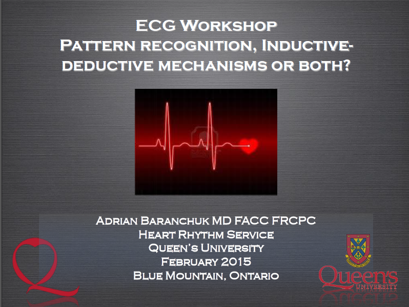
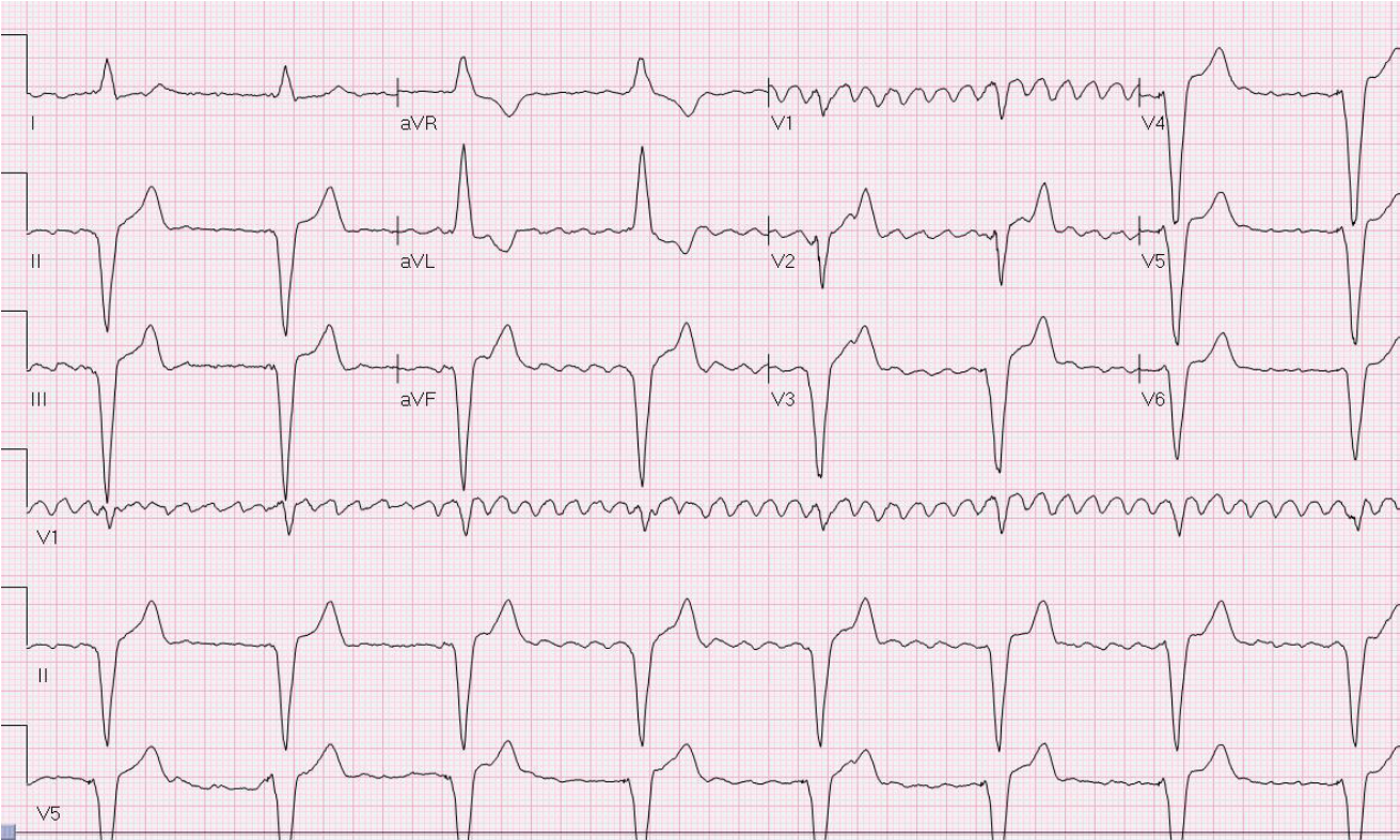
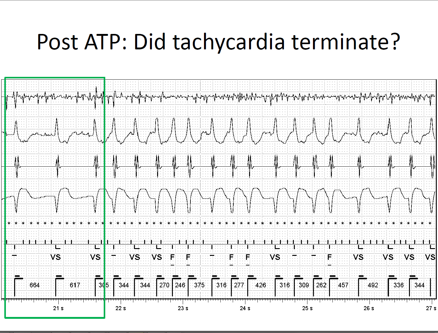
ECG Workshop by Dr. A. Baranchuk & Dr. K. Quinn

Outline of the 11th IWAS programOutline of the 11th IWAS program

ECG in Cardiac Devices by Dr. A. Pinter

Outline of the 11th IWAS program

ICD Troubleshooting (case-based) by Dr. A. Pinter

Telemetry Workshop (level 1) by Dr. D. Kagal & Dr. I. Tiong

Telemetry Workshop (level 2) by Dr. D. Kagal & Dr. I. Tiong

Telemetry Workshop (level 3) by Dr. D. Kagal & Dr. I. Tiong

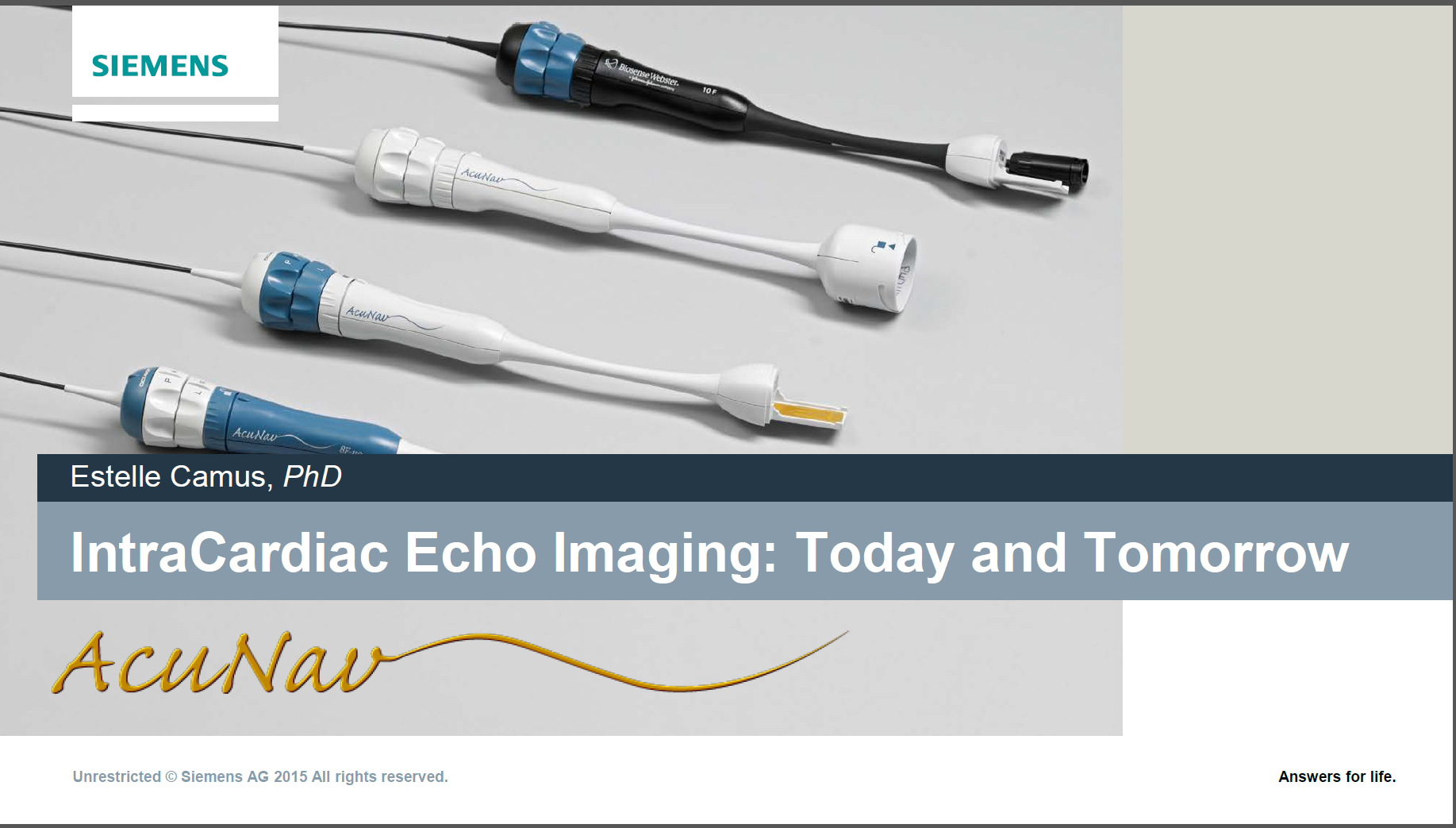
Intracardiac Echo – ICE, AcuNav by Dr. E. Camus

Managing Cardiac Devices at End of Life: Our Clinic’s Role in Proactive Care by
D. Campbell

Post Traumatic Stress in the Cardiac Patient by Dr. B. Baker

Post Implantable Defibrillator Shock Psychology Counselling by Dr. B. Baker

How to Become a Successful Researcher in Cardiology by Dr. M. Hartleib

Challenging Arrhythmia Cases by Dr. B. Makanjee

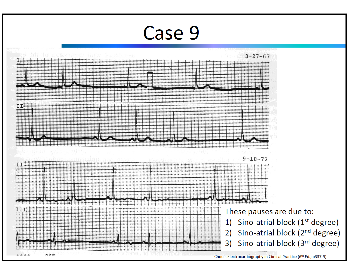
Pacemaker Troubleshooting (case-based) by Dr. D. Ng

Challenging Arrhythmia Cases by Dr. D. Ng

What is the Strategy of Ablation After STAR AF Trial by Dr. G. Amit

“Robotics in Ablation of Atrial Fibrillation” & “V-drive: Benefits and Challenges From User Perspective” by Dr. W. Spear

Indications for PVC Ablation by Dr. G. Nair

Duelling “PVI vs. Pace and Ablate” by Dr. G. Nair

Role of Stress Test in Arrhythmia Management by Dr. J. Healey

Magnet Application on “CIEDs” by Dr. D. Yung

Substrate Mapping Techniques and Technologies by Dr. D. Burkhardt

Robotics for Ischemic/structural VTs: Practical Tips and Tricks by Dr. D. Burkhardt

Post Implantation Wound Care by S. Turner

Duelling “Antiarrhythmic vs. Ablation of VT” by Dr. D. Burkhardt

Myths and Facts: A Discussion of MRI Imaging for Pacemaker and ICD Patients by M. Reitzel

Best Abstract Presenters

First Place went to Fariha Sadiq Ali for “Advanced Interatrial Block Predicts New Onset Atrial Fibrillation in Patients with Severe Heart Failure and Cardiac Resynchronization Therapy”.

F_Sadiq_Ali_tb

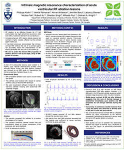
Second Place went to Philippa Krahn for “Intrinsic Magnetic Resonance Characterization of Acute Ventricular RF Ablation Lesions”.

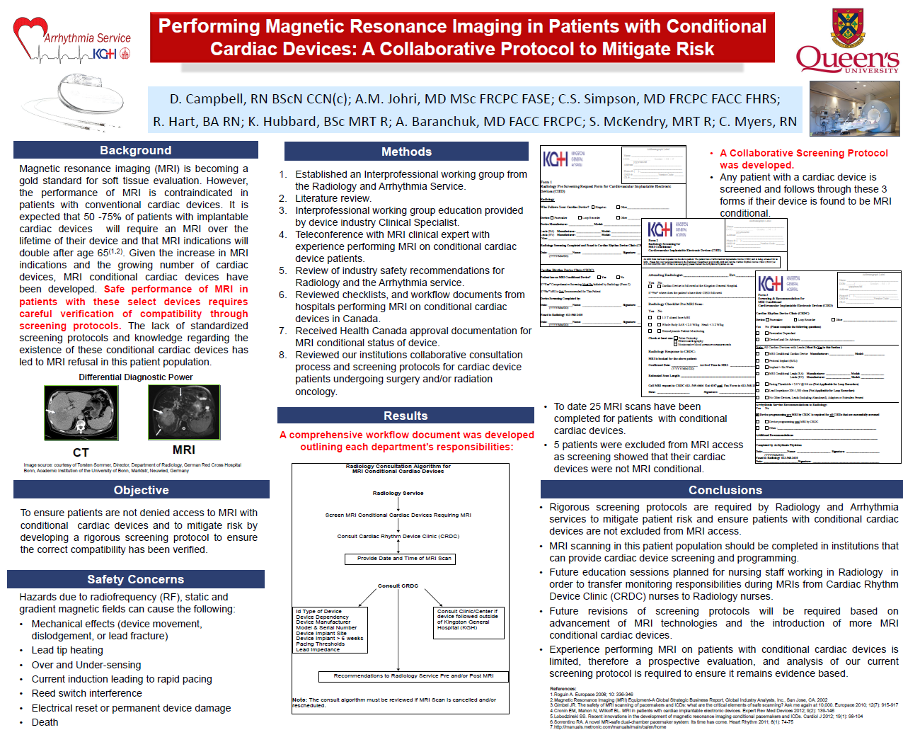
Third Place went to Debra Campbell for “Performing Magnetic Resonance Imaging in Patients with Conditional Cardiac Devices: A Collaborative Protocol to Mitigate Risk”.

Photos


  • Home
  • Contact Us
  • Facebook
  • Twitter
  • Instagram

Copyright © 2025 · Altitude Pro on Genesis Framework · WordPress · Log in